Facilities
KMC is a professional management discipline focused on the efficient and effective delivery of logistics and other support services related to real property. It encompasses multiple disciplines to ensure functionality, comfort, safety, and efficiency of the built environment by integrating people, places, processes, and technology
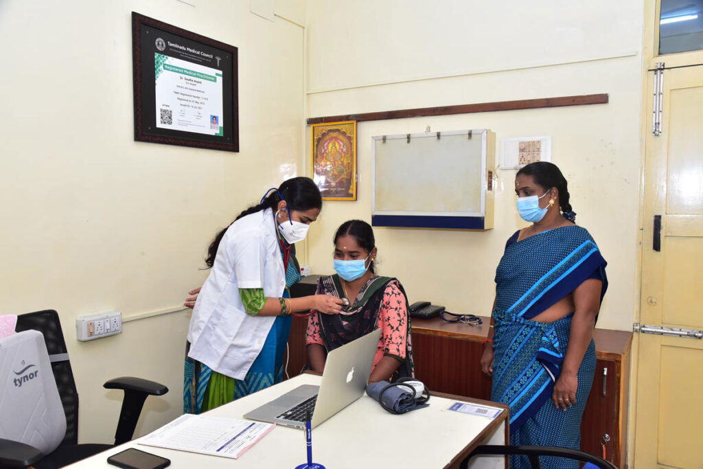
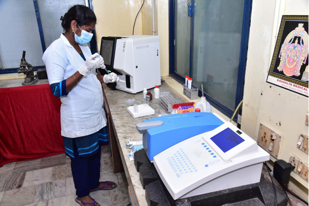
KMC is a place with controlled environments where experiments, measurements, and scientific or technological research can be done. All resources are available in our laboratory
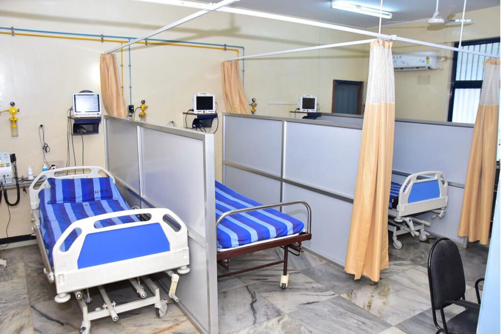
Patients in intensive care units have serious or life-threatening illnesses or injuries that require round-the-clock attention, close monitoring by life support machinery, and medicines to maintain normal bodily functioning. Highly skilled medical professionals, nurses, and respiratory therapists who specialise in caring for critically ill patients work in these facilities.
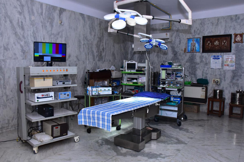
Operating rooms at the KMC Hospital are geared to handle any type of surgery. According to worldwide infection control standards, every theatre has a unique OT flooring and a dedicated air handling unit to match the air exchanges. All operating rooms have defibrillators, warmer machines, multipara monitors, LED OT lights, nice OT tables, diathermy machines, anaesthesia workstations, central suction, all medical gases, and well-trained nursing and paramedic staff.
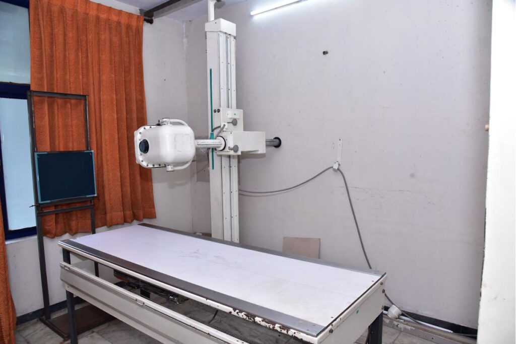
A type of electromagnetic radiation with a very low wavelength that may pass through different solid material thicknesses and behave similarly to light when it comes to film. An X-ray image was captured on camera.
Although CCTV networks are frequently employed to identify and discourage criminal activity as well as to record traffic violations, they have further applications.
For security purposes, security cameras are able to monitor areas that are not easily accessible.
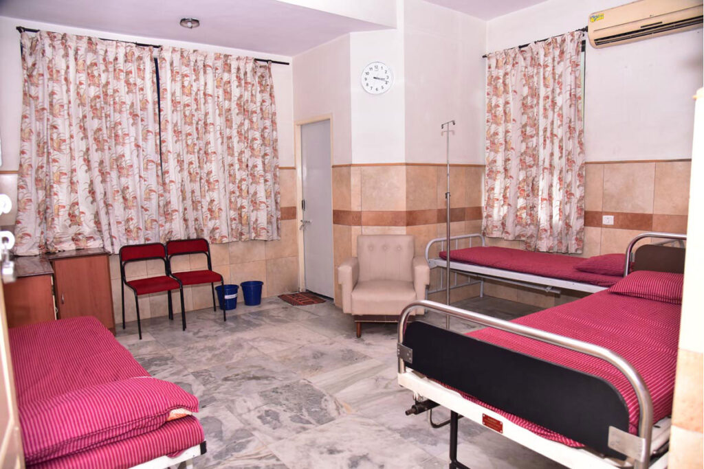
KMC provides equipment or services that are for patients, and all rooms have private facilities with private bathrooms and toilets. We have a deluxe room with an Android TV and AC.
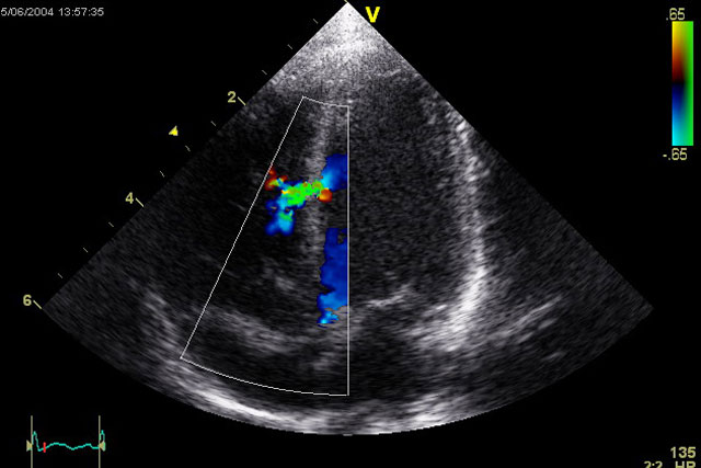
An ultrasound of the heart is known as echocardiography, echocardiogram, cardiac echo, or just an echo. Using either regular ultrasonography or Doppler ultrasound.
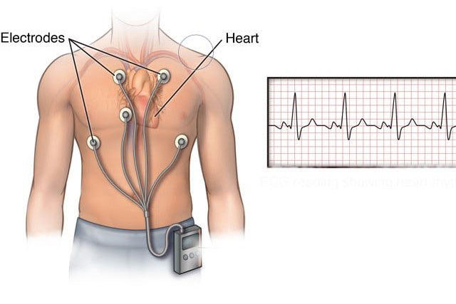
Making an ECG, a recording of the electrical activity of the heart is the procedure of electrocardiography. Using electrodes positioned on the skin, an electrogram of the heart is created, which is a graph of voltage against a time of the electrical activity of the heart.
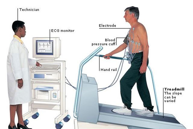
How far your heart can go before developing an irregular rhythm or losing blood flow to the heart muscle can be determined via a treadmill test (TMT) or cardiac stress test. Your doctor may want to know how your heart reacts when pushed.
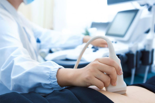
An ultrasound is a type of imaging test that employs sound waves to produce a sonogram, or image, of the organs, tissues, and other internal body components. Ultrasounds don’t use radiation like x-rays do.
